Recently, Dr Yilei Mao and his team developed a new modelling method employing three-dimensional (3D) bioprinting technology and created hepatorganoids using HepaRG cells, which preserve liver function and lengthen the longevity of mice with liver failure following abdominal transplantation. In this research, Dr Mao and his colleagues explained hepatorganoids, a model of the liver created by bioprinting HepaRG cells in three dimensions (3D), and look at how it affects both in vitro and in vivo liver function.
Liver cancer is one of the most fatal tumours globally, with five-year overall survival rates of 18% in the US and 12.1% in China. Hepatocellular carcinoma (HCC) is the most frequent form of liver cancer, accounting for 90% of all liver cancers. Patients with advanced HCC are being treated of choice with empirical systemic therapy. However, because of their heterogeneous character, the effectiveness of different systemic medications for advanced HCC patients differs greatly across people. This work intends to overcome the shortage of reliable and accessible in vitro models for patient-specific medication screening for HCC. Here, researchers developed a customized model for hepatocellular carcinoma by extending the modelling approach. Dr Mao and his colleagues in Beijing are the pioneers of this approach because they developed 3D bioprinted hepatorganoids (3DP-HOs) for prospective liver function replacement and primary HCC personalized models (3DP-HCCs) for HCC precision therapy. Even though their research is still in its early stages, it is excellent news for the industry.
A Potential Liver Function Replacement Tool: 3DP-HOs
The primary successful therapy for people with end-stage liver disease or acute liver failure is liver transplantation. The lack of organ donors, however, is a significant obstacle to treating end-stage organ failure. According to the Health Resources & Services Administration, 106,075 men, women, and children were waiting for organ transplants in the United States as of June 10. However, the average number of organs donated by live people each year is only around 6,000, while the average number of organs donated by dead people each year—around 8,000—is 3.5. This situation calls for liver function replacement therapy, which may be utilized as supportive treatment till liver transplantation (bridge-to-transplant) or liver regeneration (bridge-to-recovery).
Numerous attempts have been done for years to produce functioning cells in vitro for possible use in replacing liver function. The spatial organizing of these cells into constructions of useful tissues or organs has, however, remained a significant challenge. The function of the liver cells in vitro was virtually hard to sustain, according to many methods.
Even after years of bioprinting technological advancement, it is still difficult to properly create different functioning organs using 3D printing and use them in regenerative medicine. The medical needs of the growing number of patients with end-stage liver disease cannot be met by limited orthotropic liver transplantations, partial extracorporeal hepatic support, or small-scale hepatocyte injection therapy. Numerous initiatives have been launched for years to produce in vitro functioning cells for possible application in regenerative medicine. The spatial organizing of these cells into constructions of useful tissues or organs has, however, remained a significant challenge. An adequate cell source with a well-organized structure for further manipulation during implantation, a 3D microenvironment with efficient mass transfer to maintain liver-specific functions that closely resemble those in vivo, and ease of handling and immobilization during transplantation are important factors to consider when building a reliable hepatic organoid in vitro for implantation.
People are thrilled that Dr Mao’s team has just had some success. Three-dimensional (3D) bioprinting technology was utilized to create liver tissue models known as “hepatorganoids,” or “3DP-HOs,” utilizing a human liver progenitor cell line and a biocompatible scaffolding material.
By 3D bioprinting human liver tissues, Dr Mao and his colleagues were able to extend the lives of mice with liver failure. HepaRG is a regularly used hepatic progenitor cell line. They provided a proof of concept for the use of 3D bioprinted human liver tissues as organ donors and explained the potential benefits of 3D bioprinting technology in liver function replacement treatment. The study was intriguing because of its many potential uses. This technique may be used to augment substantial hepatectomy resection and interim maintenance of the liver during a waiting time for transplantation, or it may be used as a rescue bridge to move from liver failure to liver regeneration. Additionally, in clinical transplantation, the technology may be combined with the proper cells to build artificial organs that can be implanted into the body, perform typical physiological activities in vivo, and provide cutting-edge treatments for certain disorders. A long-term objective is to create a transplantable liver organ using 3D bioprinting for clinical treatment. But Dr Mao and his team were able to achieve a tremendous improvement. The technology related to in vitro manipulation of cultured human hepatocytes is anticipated to significantly aid in the creation of improved clinically transplantable 3D bioprinted liver tissues or organs in the future, which in turn will aid in the development of new clinical methods to treat liver diseases.
A 3D Bioprinted Primary Liver Cancer Model for Precision Treatment
Hepatocellular carcinoma (HCC), the most prevalent kind of liver cancer, claims more than 1200000 lives annually, making it one of the most deadly malignancies in the world. Patients with advanced HCC are being treated of choice with empirical systemic therapy. However, there are considerable individual differences in the effectiveness of several systemic medications for patients with advanced HCC. Therefore, the requirement for patient-specific therapy is critical.
In his State of the Union Address on January 20, 2015, President Barack Obama put forward a precision medicine strategy. According to common understanding, precision medicine employs diagnostic techniques and therapeutic regimens tailored to each patient’s requirements in light of their unique genetic, biological, or psychological traits. To provide patients with precise care, Dr Mao and his team have tried several methods, including genetic testing. Targeted treatment is, however, mostly ineffective due to its poor efficiency, even when genetic testing is used to identify the precise targets. Sorafenib, for instance, has an objective response rate (ORR) of less than 3%, but regorafenib has an ORR of 11%. The ORR for lenvatinib, which is just 24%, is the best of any first- or second-line systemic therapy. As a result, in vitro tumour model research is receiving more attention.
Six individuals had surgery, and six had HCC specimens taken. To create the bioink for printing, primary HCC cells were separated and combined with sodium alginate and gelatin. After that, three-dimensional bio-printed HCC models made from patient cells (called 3DP-HCCs) were effectively created and thrived well under long-term culture. These models maintained the characteristics of the original HCCs, including stable biomarker expression, steady preservation of the genetic changes, and stable expression patterns. Drug screening data may be presented mathematically and intuitively using 3DP-HCC models.
Using primary human hepatocellular carcinoma cells for 3D bioprinting, we generated 3DP-HCC models with success. Our findings demonstrated the benefits of the 3DP-HCC model, which included retaining the biological and genetic characteristics of the original tumour, being established quickly and simply, obtaining uniformity in establishment with consistent cell density and even cell distribution, being independent of the intrinsic proliferative capacity of the original tumour, being able to be adjusted and rewritten through flexible programs during the 3D-printing process, and being capable. As a result, 3DP-HCC models will be able to function as a trustworthy in vitro model system for screening various potential medications for the individual treatment of HCC patients.
Following are the Benefits explained by Dr Mao’s team for the 3DP-HCC model
l Maintaining the biochemical and genetic characteristics of the original tumour; being created through simple, rapid processes
l To achieve uniformity in establishment with consistent cell density and dispersion, and to be independent of the inherent proliferative potential of the initial tumour via adaptable programming, be modifiable and rewriteable throughout the 3D printing process; capable of long-term culture and becoming the desired models with various parameters.
As a result, 3DP-HCC models will be able to serve as a trustworthy in vitro model system for evaluating several candidate medications for patients with HCC who want individualized care.
About the Author

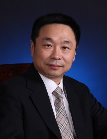
Professor of Surgery Yilei Mao works at the Department of Liver Surgery at Peking Union Medical College (PUMC) Hospital, Chinese Academy of Medical Sciences, and PUMC. He is Deputy Chair of the Chinese Society of Liver Surgeons and a Ministry of Health expert committee member for National Tumor Standardized Diagnosis and Treatment (Liver Cancer). He belongs to the American Society of Nutrition. He earned his PhD in 1997 from Lund University, Sweden, after graduating domestically and residency training in Australia.
Dr Mao received post-doctoral and clinical training at Harvard Medical School and Massachusetts General Hospital’s Surgical Oncology. Dr Mao received the 2001 Li Foundation Heritage Prize for originality, three national Excellent Researches of General Surgeons honours, and the 2011 China National Cancer Center award. He leads various China Medical Board of New York and National Natural Science Foundation-funded research initiatives. He has almost 100 journal papers. Dr Mao founded Hepatobiliary Surgery and Nutrition and is its editor-in-chief (IF: 8.26). He elevated the journal to a top surgical publication in 10 years.
Media Contact
Company Name: Index of Sciences Ltd
Contact Person: Stella Richards
Email: Send Email
Phone: +441442781196
Address:Kemp House 160 City Road
City: London
State: England
Country: United Kingdom
Website: https://gut.bmj.com/content/70/3/567
